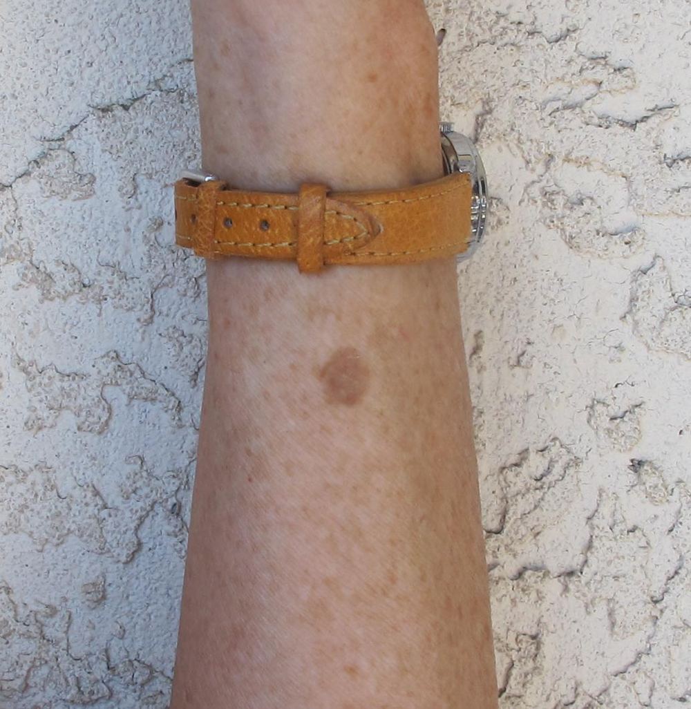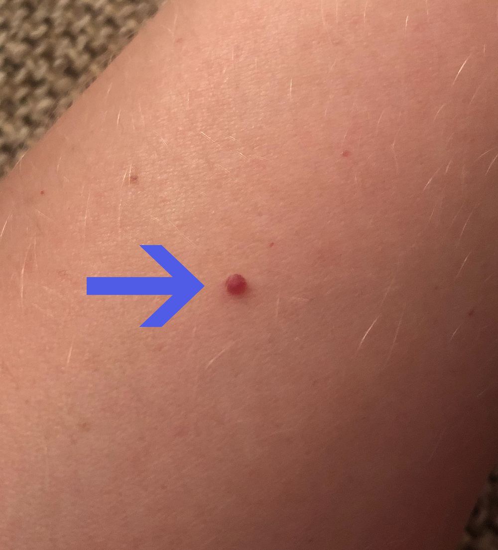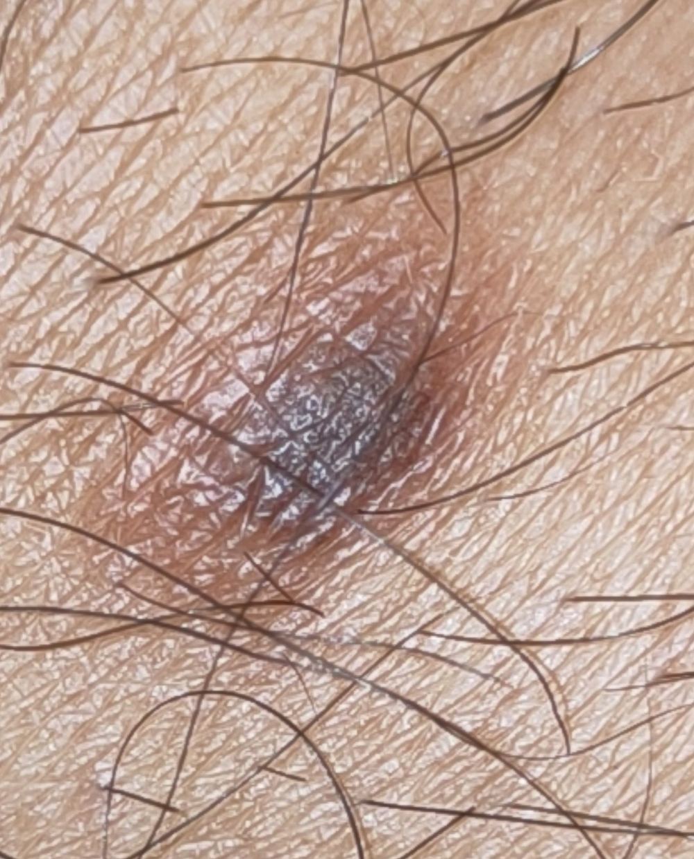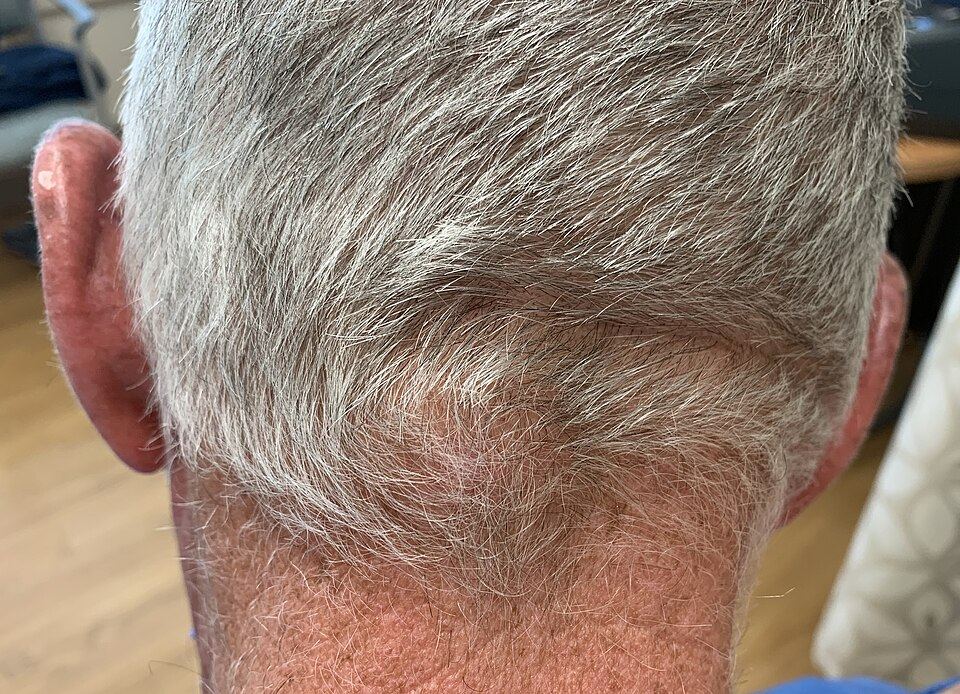
What a patient may notice
- A new spot or an existing mole that changes
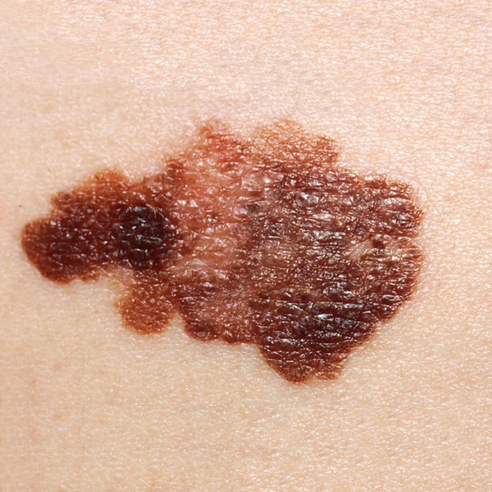
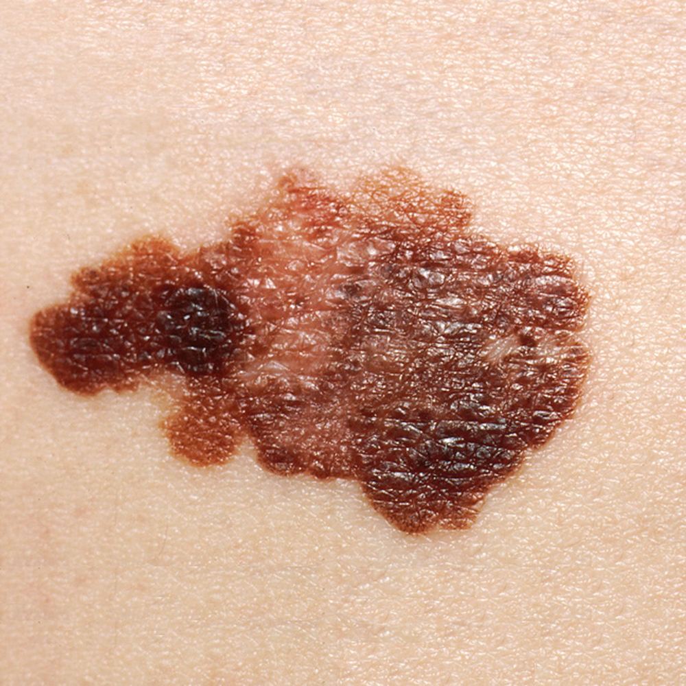
- Asymmetry, irregular border or several colours
- A firm, raised lesion that is growing
- Bleeding, itching or ulceration without another explanation
Potentially serious · early review matters
Melanoma
A cancer of pigment-producing melanocytes. It is less common than BCC or SCC but is more likely to spread if not found early.
How common?
Melanoma is one of Australia’s most commonly diagnosed cancers. Risk rises with age, but it can occur in younger adults.
Main causes and risks
UV exposure—especially episodes of sunburn—is the major preventable risk. Fair skin, many or atypical moles, family or personal history, and immune suppression also increase risk. Melanoma can occur on skin with little sun exposure.
Main clinical types
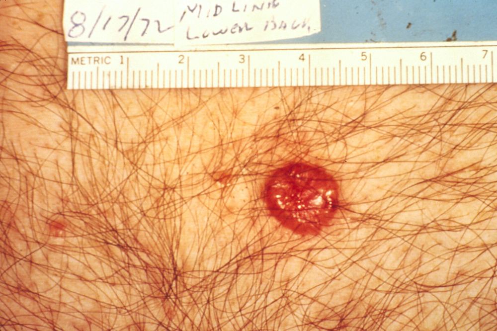
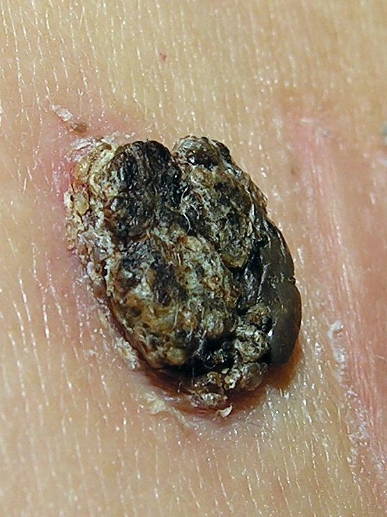
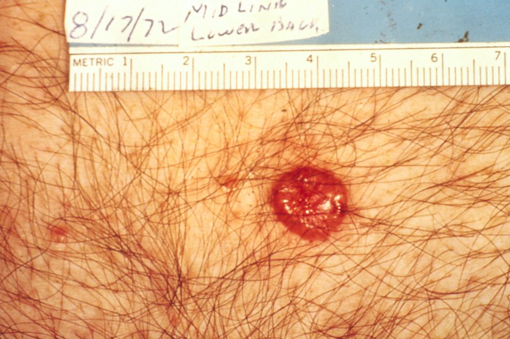
Superficial spreading melanoma commonly enlarges across the skin before invading more deeply. Nodular melanoma often presents as a firm, rapidly enlarging lump and may not satisfy the ABCDE rule. Lentigo maligna melanoma usually develops on chronically sun-damaged facial skin in older adults. Acral lentiginous melanoma occurs on palms, soles or beneath nails and is relatively more important in darker skin types. Amelanotic melanoma may be pink, red or skin-coloured.
What is melanoma in situ?
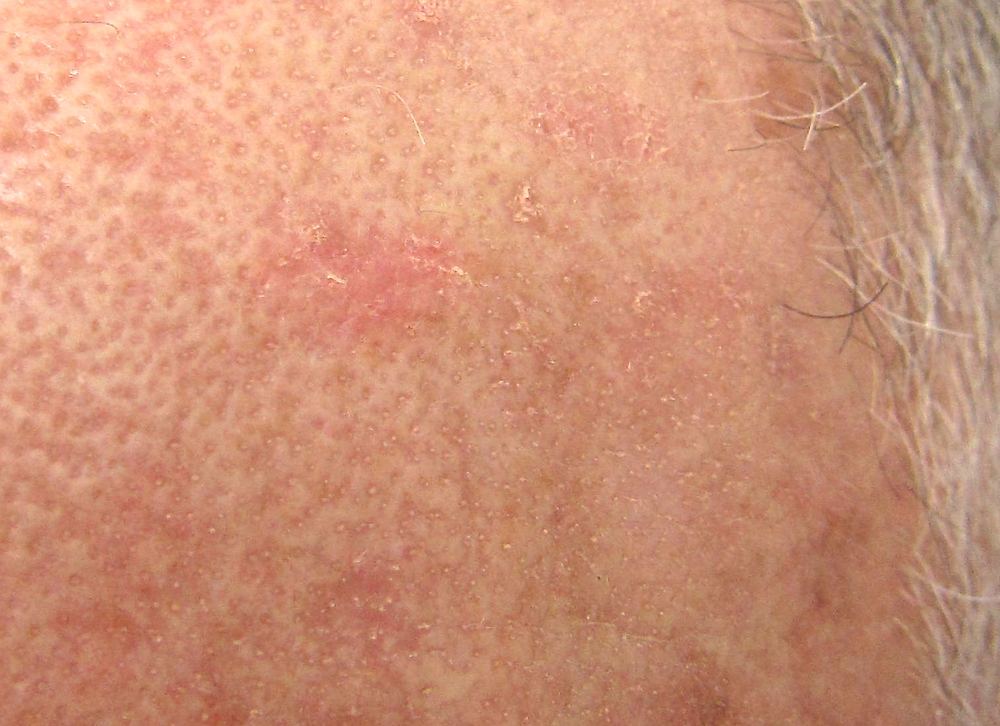
Melanoma in situ (Stage 0 melanoma) means abnormal melanocytes are confined to the epidermis—the outer layer of skin—and have not invaded the dermis. It often appears as a slowly enlarging, irregular flat patch with uneven colour; lentigo maligna is an in-situ subtype usually found on chronically sun-damaged facial skin. Pathology confirms the diagnosis. Because there is no dermal invasion, Breslow thickness is not assigned and the lesion itself cannot spread to lymph nodes or distant organs. Complete surgical removal is usually curative, commonly with a further margin after the diagnostic biopsy. Large or poorly defined facial lesions may need staged margin-controlled surgery. Untreated lesions can enlarge and some eventually develop an invasive focus, so treatment and ongoing skin surveillance remain important.
What is desmoplastic melanoma?
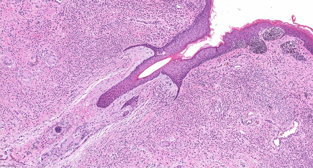
Desmoplastic melanoma is a rare invasive melanoma in which malignant spindle-shaped melanocytes lie within dense fibrous tissue. It usually affects older, fair-skinned people on chronically sun-damaged head or neck skin and may arise beneath lentigo maligna. It may look like a firm, pale, pink or scar-like thickening rather than a dark mole, making recognition and partial-biopsy diagnosis difficult. Involvement of small nerves (neurotropism) can cause pain, tingling or numbness and increases concern for local recurrence. Diagnosis requires an adequately deep biopsy and specialist dermatopathology. Wide surgical excision is the main treatment; margin-controlled surgery, radiotherapy and staging investigations are considered according to site, thickness, margins, neurotropism and whether pathology describes a pure or mixed subtype. Management should be planned by an experienced melanoma team.
Appearance and usual sites
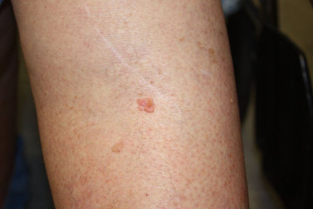
Melanoma may be a new lesion or a changing existing mole. Warning patterns include asymmetry, an irregular edge, uneven colour, evolution, or a spot unlike the person’s other lesions. Men more often develop melanoma on the trunk and women on the legs, but it can arise anywhere—including scalp, nails, soles, genital skin and rarely the eye or mucosa.
What may be seen with dermoscopy?
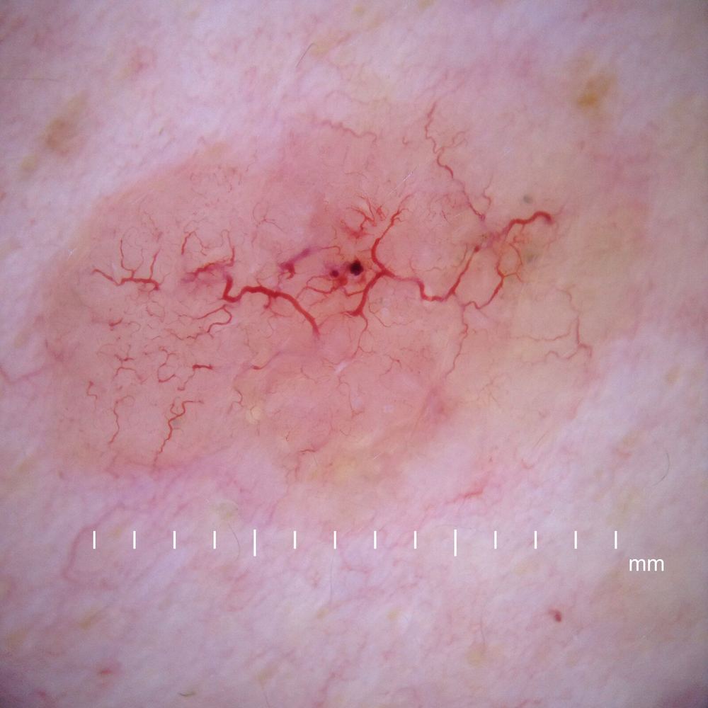
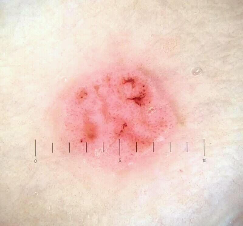
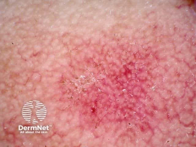
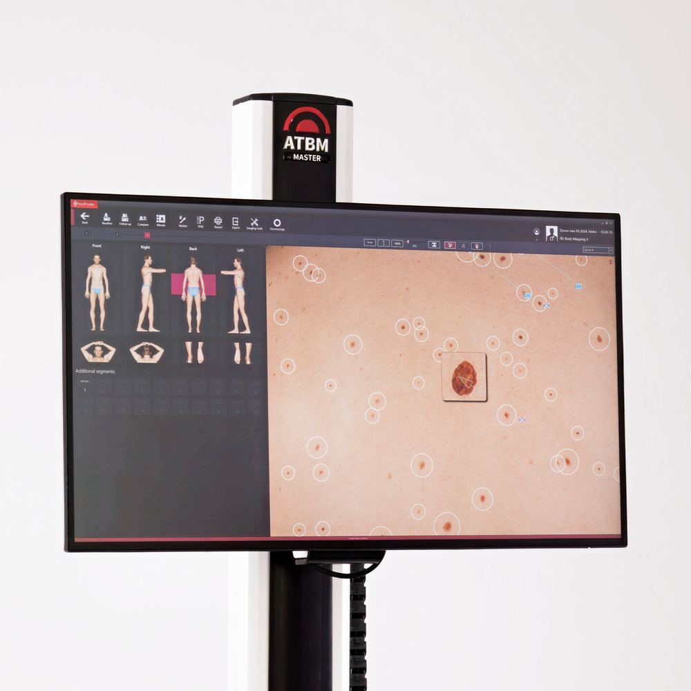
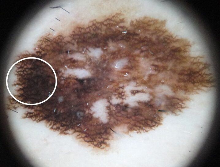
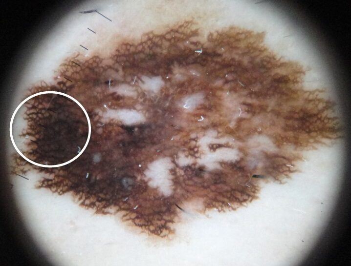
A trained clinician may see an atypical or broadened pigment network, irregular dots or globules, streaks, blue-white structures, regression, asymmetrical structureless areas or atypical blood vessels. No single feature proves melanoma; the complete pattern and clinical history matter.
What else can resemble it?
Atypical or irritated moles, seborrhoeic keratosis, pigmented BCC, dermatofibroma, a blood blister and benign nail pigmentation may resemble melanoma. Conversely, melanoma can look deceptively harmless, so persistent change should be examined rather than matched to photographs.
How is it diagnosed?
Assessment includes history, examination of the lesion and surrounding skin, comparison with other moles and usually dermoscopy. When feasible, the entire suspicious lesion is removed with a narrow margin for pathology. The report records features such as melanoma thickness and ulceration. A wider excision is then planned; sentinel lymph-node biopsy and scans are considered only for selected stages.
Treatment options
Surgery is the main treatment for localised melanoma: diagnostic excision is followed by a wider excision based on pathology. Selected patients may be offered sentinel-node biopsy. Higher-risk or advanced melanoma may require specialist immunotherapy, targeted therapy when an actionable mutation is present, radiotherapy in selected situations, or a combination planned by a multidisciplinary team.
Overall outlook
Outlook is excellent for many thin, early melanomas. It becomes less favourable once melanoma is thicker or has spread, which is why prompt assessment of change matters.
After treatment
Follow-up is individualised according to stage and personal risk. It commonly includes scar and lymph-node review, full-skin examinations, self-check education and lifelong sun protection. A previous melanoma increases the chance of another melanoma and other skin cancers.